Coronary Calcium Found on a Chest CT
Review date: 2026-09-17 · Credential: 110*********650
Abstract
Coronary Calcium Found on a Chest CT
Coronary Calcium Found on a Chest CT: How to Verify Next Steps
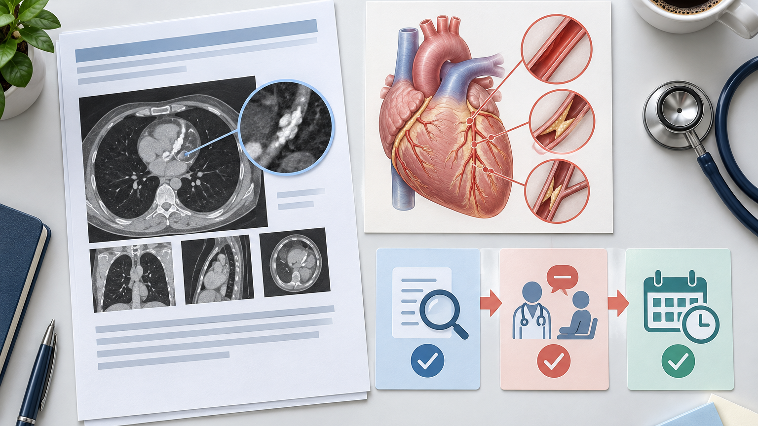
A "coronary calcium" note on a chest CT report is often an incidental finding when examining the lungs, mediastinum, or other chest structures. It suggests possible calcific changes in the coronary artery walls, but by itself, it usually cannot determine the degree of vessel narrowing or confirm the need for medication or interventional treatment.
The next step is not to self-diagnose but to verify the report and original images, consider symptoms and overall cardiovascular risk, and have the results interpreted by appropriate medical professionals. This article is for general medical education and does not replace diagnosis or individualized treatment advice.
Clarify the task first: incidental findings need review, not equivalent to having had a heart attack
The coronary arteries supply blood to the heart muscle. Coronary calcium seen on imaging typically refers to calcium deposits within the coronary artery walls. It can be a piece of information for assessing atherosclerotic risk, but its clinical significance depends on the extent of calcification, imaging method, age, symptoms, and other risk factors.
When seeing this description, first distinguish several concepts:
Coronary calcium is not the same as an ongoing myocardial infarction.
Seeing calcium does not confirm severe stenosis.
The absence of calcium in the report does not rule out coronary artery disease.
More prominent calcium may indicate higher long-term cardiovascular risk, but a complete evaluation is still needed.
The need for further testing or treatment cannot be determined by a single word in the report.
If there are no acute symptoms, you can usually organize the information and arrange for a review. If there are persistent or worsening chest discomfort, significant shortness of breath, or other warning signs, do not wait for a routine follow-up.
Find the calcium description, examination date, and original image information from the report
First save the complete report; do not only keep a screenshot containing "coronary calcium." When reviewing the report, record the following information:
Examination date and facility.
Whether the examination used ECG gating.
Whether intravenous contrast was used.
How the report describes the calcium, e.g., mild, moderate, severe, scattered, or extensive.
Whether specific vessel locations are mentioned.
Whether the heart, aorta, lungs, or other chest abnormalities are also described.
Whether a formal coronary calcium score is provided.
Whether the original images are accessible via image disc, patient portal, or other compliant means.
The severity description in a routine chest CT report may be informative, but different scanning protocols and reporting styles vary. Do not interpret "mild" or "small amount" as no risk, nor interpret "severe" or "extensive" as requiring surgery.
When visiting a doctor, provide both the report and original images. The doctor may need to confirm whether the imaging findings are indeed coronary calcium, whether the description is complete, and whether this examination can serve the purpose of subsequent evaluation.
Why a non-cardiac chest CT cannot directly replace a full risk assessment
CT scans for lung or other chest issues are not necessarily performed according to coronary assessment requirements. The heart is beating; scan parameters, slice thickness, ECG gating, and contrast use can all affect the visualization of the coronary arteries.
Therefore, a non-cardiac chest CT can suggest the presence of calcium but often cannot independently answer the following questions:
Whether and to what degree the coronary lumen is narrowed.
Whether a particular calcification is causing current symptoms.
Whether non-calcified plaques are present that are difficult to identify on such scans.
Whether myocardial blood supply is affected.
Whether medication, coronary intervention, or surgery is needed.
Formal coronary calcium scoring, coronary CT angiography, and other cardiac tests have different purposes, and not all are necessary after detecting calcium. The choice of test also depends on symptoms, baseline risk, renal function, contrast-related risks, radiation exposure, heart rate, and previous test results. The specific plan should be decided by a clinician based on the evaluation purpose.
Organize chest symptoms, smoking history, family history, and previous tests before the visit
Imaging results are best interpreted when placed in the context of personal risk. Before the visit, prepare a concise record.
Regarding symptoms, record the presence or absence of chest pain, chest tightness, pressure, shortness of breath, palpitations, unusual fatigue, or reduced exercise tolerance, and note:
When they started.
Whether they appear suddenly or gradually.
How long each episode lasts.
Whether they are triggered by walking, climbing stairs, emotional stress, or cold.
Whether they are relieved by rest.
Whether they are accompanied by sweating, nausea, dizziness, or discomfort radiating to the arms, shoulders, back, neck, jaw.
Also gather information that may affect cardiovascular risk assessment:
Current and past smoking status.
History and recent results related to blood pressure, lipids, and glucose.
Known heart disease, cerebrovascular disease, or peripheral arterial disease.
Whether first-degree relatives had early-onset coronary heart disease or related cardiovascular events.
Daily activity level and recent changes.
Current prescription medications, over-the-counter drugs, and supplements.
Previous ECG, echocardiogram, stress test, coronary imaging, or chest CT results.
The definition of "early" in family history and the weight of a particular risk factor should be judged by a physician in context. Do not assume you have the same disease just because a family member has it, nor ignore current symptoms because of no family history.
Review question table: extent of findings, risk background, and purpose of subsequent evaluation
During the visit, you can communicate with the doctor using the following questions:
Review aspect | Questions to ask | Misconceptions to avoid |
|---|---|---|
Imaging findings | Where is the calcium, how to interpret the severity description, is a re-read needed | Equating a descriptive word directly to percent stenosis |
Test capability | What protocol was used for this chest CT, what questions can it answer, what can it not answer | Assuming all chest CTs can fully evaluate the coronary arteries |
Risk background | How do symptoms, blood pressure, lipids, glucose, smoking history, and family history affect the overall judgment | Looking only at imaging and ignoring clinical risk |
Purpose of follow-up | If further testing is recommended, what clinical question is it meant to answer | Repeating tests just to "be thorough" |
Management plan | Whether lifestyle changes, repeat labs, or medication are needed, and when to follow up | Starting, stopping, or changing medication doses on your own |
If medication is recommended, confirm the indication, expected effect, common side effects, contraindications, interactions with current medications or supplements, and what to monitor. Aspirin, lipid-lowering drugs, etc., are not to be started or stopped based solely on one imaging term.
Which current symptoms require immediate help rather than waiting for report review
If symptoms possibly indicating an acute cardiovascular problem appear, contact local emergency services or seek urgent medical evaluation immediately, rather than waiting for a routine clinic visit or driving yourself.
Warning signs include:
New, persistent, unrelieved, or rapidly worsening chest pain, tightness, pressure, or squeezing.
Chest discomfort with significant shortness of breath, cold sweat, nausea, vomiting, dizziness, or fainting.
Discomfort radiating to one or both arms, shoulders, back, neck, jaw, or upper abdomen.
Sudden severe fatigue, altered consciousness, or a sense of impending doom.
Patients with known cardiovascular disease experiencing symptoms that are different or clearly worse than usual.
Acute cardiac symptoms can be atypical in different people; some may present mainly with shortness of breath, unusual fatigue, nausea, or upper abdominal discomfort. Whether it is an emergency cannot be determined by an online article. When in doubt, have emergency or medical professionals assess promptly.
Verification boundaries: cannot judge stenosis severity or treatment need based on a single imaging term
Calcium is part of the risk information, not a complete diagnosis. The amount of calcium may correlate with coronary atherosclerotic burden, but there is no simple one-to-one correspondence between calcium appearance and lumen stenosis. Routine chest CT can also be affected by cardiac motion, scan coverage, and image quality.
Therefore, the following practices are unreliable:
Self-judging that a particular vessel is blocked based on the word "calcium" in the report.
Using the presence or absence of chest pain to confirm or exclude coronary artery disease.
Interpreting an incidental finding as certain to cause a heart attack in the near future.
Comparing adjectives across reports without considering scanning protocols and image differences.
Taking aspirin or other medications on your own out of fear of calcium.
Permanently shelving the result because there are no symptoms, without further risk factor assessment.
Follow-up evaluation may include history taking, physical examination, and blood pressure, lipids, or glucose information. Whether additional cardiac testing is needed depends on whether it can answer a clear clinical question and change management. Tests themselves carry burdens such as radiation, contrast reactions, incidental findings, and further workup, so more is not always better.
Final actions: save report and images, and confirm who is responsible for interpreting results
After detecting coronary calcium, you can proceed in the following order:
Save the complete report, examination date, and how to obtain original images.
Review the original report text; do not rely on paraphrases or partial screenshots.
Organize current symptoms, risk factors, medications, and previous tests.
Confirm which doctor is responsible for interpreting the results and arranging follow-up, starting with the doctor who ordered the chest CT or the one managing your cardiovascular risk.
Ask whether image re-review, additional risk assessment, or testing with a clear purpose is needed.
Do not start, stop, or adjust prescription drugs or medications that may increase bleeding risk without medical evaluation.
If acute warning signs appear, contact local emergency services immediately.
The value of an incidental imaging finding is to prompt targeted verification and risk management. Giving the report, original images, symptoms, and personal risk information to a clearly identified result interpreter is more helpful for forming a safe, reasonable next step than speculating about the words "coronary calcium" alone.